Isolation and Immunological Detection of Mycobacterium Tuberculosis from HIV and Non-HIV Patients in Benue State, Nigeria.

Isolation and Immunological Detection of Mycobacterium Tuberculosis from HIV and Non-HIV Patients in Benue State, Nigeria.

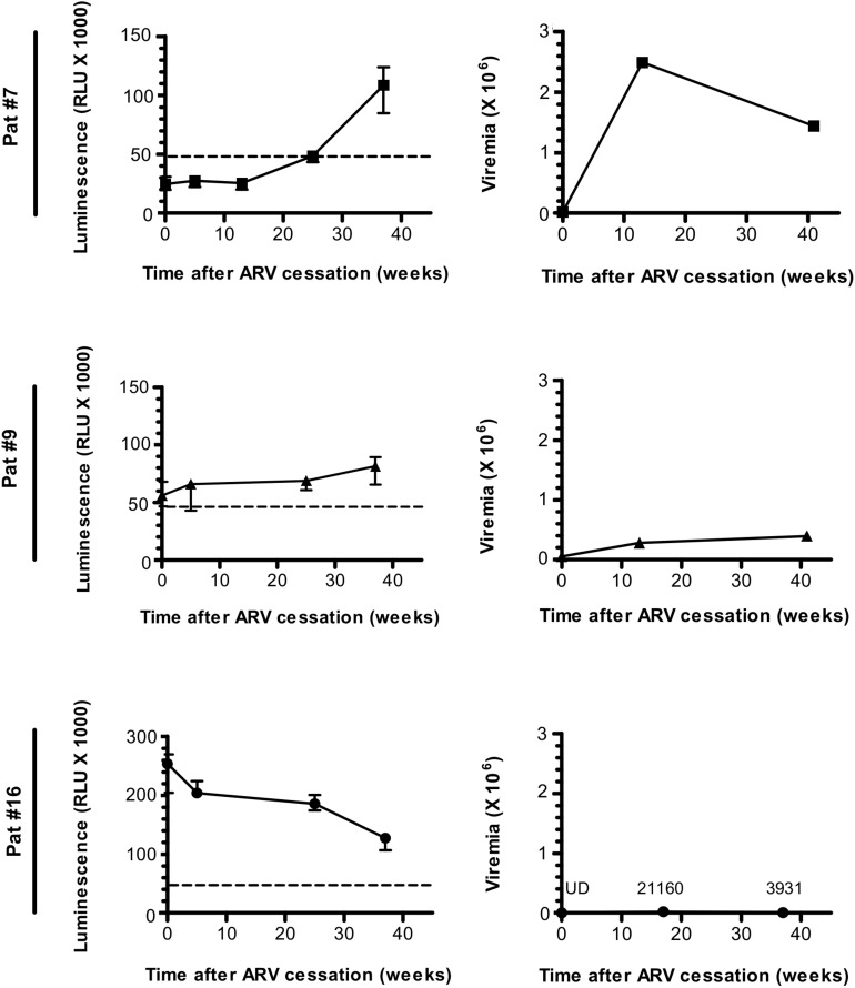
Immunological methods are essential instruments for tuberculosis epidemiology; though its use is underutilized in Nigeria. In this research, we report the epidemiological outlook of Mycobacterium tuberculosis amongst HIV sufferers in Benue State, Nigeria.

Sputum samples had been collected from 425 suspected TB sufferers from July 2016 to February 2018 and subjected to acid-fast microscopy, GeneXpert MTB/RIF, processed utilizing NALC-NaOH and cultured on Lowenstein-Jensen media. The isolates obtained had been recognized by SD-Bioline® assay.

ResultsThe prevalence of TB by acid-fast microscopy was 35(15.9%). The prevalence of TB by acid-fast bacilli was considerably (χ2 = 8.458; P = 0.003) highest among the many 15-34 years age group (22.0%) in contrast with different age teams. TB prevalence was considerably (χ2 = 4.751; P = 0.029) greater amongst sufferers from rural areas than these from city middle (23.8% vs 14.1%).

GeneXpert assay detected 64(15.1%) TB circumstances of which sufferers from rural areas had considerably (χ2 = 8.104; P = 0.017) greater prevalence of TB than sufferers from city areas (23.8% vs 12.9%). The total rifampicin resistance TB was 3.1%. Also, sufferers from rural areas had considerably (χ2 = 10.625; P = 0.005) greater rifampicin resistance in contrast with affected person from city areas (8.3% vs 1.3%). Of the 126(29.7%) mycobacterial isolates, 42(33.33%) had been recognized as MTBC and 84 (66.67%) as NTM by SD-Bioline® assay.

The research revealed that Mycobacterium tuberculosis an infection remains to be a significant public well being drawback, with comparatively excessive prevalence charge of rifampicin resistance amongst HIV optimistic sufferers. Further research are wanted for early detection and remedy intervention needed for an infection management.

Isolation and Immunological Detection of Mycobacterium Tuberculosis from HIV and Non-HIV Patients in Benue State, Nigeria.

Fulminant central nervous system varicella-zoster virus an infection unexpectedly recognized by metagenomic next-generation sequencing in an HIV-infected affected person: a case report.

Varicella-zoster virus (VZV) an infection might be recognized clinically as soon as classical rash happens however the prognosis is difficult when typical rash is absent. We reported a case of fulminant central nervous system (CNS) VZV an infection in a human immunodeficiency virus (HIV)-infected affected person with out typical VZV-related rash. CNS VZV an infection was surprising recognized by metagenomic next-generation sequencing (mNGS).A 28-year-old HIV-infected affected person introduced with neurological signs for Three days.

The affected person, who was not suspected of VZV an infection at admission, rapidly progressed to deep coma throughout the first 24 h of hospitalization. An unbiased mNGS was carried out on DNA extract from 300 μL cerebrospinal fluid (CSF) with the BGISEQ-50 platform.

The sequencing detection recognized 97,248 (out of 38,561,967) sequence reads uniquely aligned to the VZV genome, and these reads coated a excessive share (99.91%) of the VZV. Presence of VZV DNA in CSF was additional verified by VZV-specific polymerase chain response and Sanger sequencing.

Altogether, these outcomes confirmed CNS VZV an infection.This research means that mNGS could also be a helpful diagnostic device for CNS VZV an infection. As mNGS might establish all pathogens instantly from CSF pattern in a single run, it has the promise of strengthening our capacity to diagnose CNS infections in HIV-infected sufferers.

A Pilot Study of the Humoral Response Against the AntiSense Protein (ASP) in HIV-1-Infected Patients.

The existence of an antisense Open Reading Frame (ORF) that encodes a putative AntiSense Protein (ASP) on the proviral genome of Human Immunodeficiency Virus type 1 (HIV-1) was a source of debate for 30 years.

During the last years, some progresses have been made to characterize the cellular immune response against ASP in HIV-1 seropositive patients. However, no tools were available for the detection of antibodies to ASP in the plasma of HIV-1-infected patients during the natural course of the infection.

The aim of our study was to develop a Luciferase Immuno-Precipitation System (LIPS) to monitor the quantitative detection of ASP-specific antibodies in the plasma of HIV-1-infected patients [antiretroviral therapy (ART) naive-patients, patients under ART and HIV-1 controllers], patients who discontinued antiretroviral drugs (ARV). We further used this approach to delineate the epitopes of ASP targeted by antibodies.

Antibodies directed against ASP were detected in 3 out of 19 patients who discontinued ARV (15%) and in 1 out of 10 ART-naive patients (10%), but were neither detected in HIV-1 infected patients under ART nor in HIV-1 controllers. Individual variations in levels of ASP-specific antibodies were detected overtime.

Both the conserved prolin-rich motif and the core 60-189 region of ASP were found to be essential for antibody recognition in the four patients tested positive for anti-ASP antibodies, who were all untreated at the time of sampling.

Moreover, for two of these patients, increased levels of ASP-specific antibodies were observed concomitantly to viremia declines. Overall, our method may represent a useful tool to detect a humoral response to ASP in HIV-1-infected patients, which allowed us to confirm the expression of ASP during the course of HIV-1 infection. Further studies will be needed to fully characterize the humoral response to ASP in HIV-1-infected patients.

A Pilot Study of the Humoral Response Against the AntiSense Protein (ASP) in HIV-1-Infected Patients.

Comparison of two Immunoassays for Concurrent Detection of HCV Antigen and Antibodies among HIV/HCV Co-infected Patients in Dried Serum/Plasma Spots.

Hepatitis C virus (HCV) antigen/antibody (Ag/Ab) assays offer the benefit of reducing the window period compared to assays that detect only HCV-Ab. In this study the performance of the Murex Ag/Ab (Murex, Abbott) and Monolisa Ag/Ab Ultra (Monolisa, Bio-Rad) ELISAs was compared for the use of filter dried serum/plasma spots (DS/PS) with a focus on the sensitivity and the percentage of correct positive test results.

Correct positive ELISA results were assumed for samples that subsequently tested positive for HCV RNA by RT-qPCR, or RNA negative samples that tested positive in a Western blot (confirmed ELISA results).

Sensitivity was evaluated from DS/PS eluates using HCV seroconversion panels [plasma samples of subtypes-(St) 1a, 2b)] and longitudinal HCV antibody-positive serum panels (St 1b, 2b, 3a, and 4d). The proportion of correct positive test results was evaluated using 1102 newly-diagnosed HIV-positive clinical dried serum spots DSS eluates for screening of potential HCV co-infection.

For the plasma HCV seroconversion samples, which were used as a reference for DSS eluates, the Murex became reactive earlier for antigen-positive bleeds. However, for the HCV antibody-positive eluates and dilutions thereof, the Monolisa demonstrated a superior sensitivity.

Of the clinical DSS 22.8% (28/123) of samples reactive in the Murex were negative in a subsequent RT-qPCR and Western blot, while only 1.9% (2/105) of the samples reactive in the Monolisa were negative in these confirmatory assays. Our results indicate that the Monolisa provides fewer false positive results for HCV detection in DSS, whereas for undiluted plasma or serum samples, the Murex can serve as an additional diagnostic tool to narrow the window period.

Burden of tuberculosis and challenges related to screening and diagnosis in Ethiopia.

Burden of tuberculosis and challenges related to screening and diagnosis in Ethiopia.

One-third of tuberculosis (TB) circumstances in Ethiopia are lacking from look after causes that aren’t properly studied. The goal of this examine was to assess TB burden and establish challenges related to TB screening and diagnosis in Ethiopia.

A facility-based cross-sectional examine was carried out in seven well being services chosen from two areas and 2 metropolis administrations of Ethiopia utilizing stratified random sampling procedures.

The information of 1,059,065 sufferers have been included from outpatient division, HIV clinic, diabetic, and maternal-child well being clinics. Data have been collected from October to December 2018 utilizing a retrospective assessment of three years’ facility information (2015 to 2017) supplemented by a semi-structured interview with purposively chosen well being care employees and heads of the well being services.

ResultsA complete of 1,059,065 sufferers visited the well being services in three years, of these, 978,480 (92.4%) have been outpatients. Of the entire, 20,284 (2%) have been presumptive TB circumstances (with 14 days or extra cough), 12.2% (2483/20,284) of which had TB. For the kind of TB, 604 (24.3%) have been smear-positive pulmonary TB (PTB), 789 (31.8%) have been smear-negative PTB, 719 (29%) have been extra-pulmonary TB, and information have been lacking for the remaining.

TB screening was built-in into HIV clinic, outpatient division, diabetic clinic however not with the maternal and little one clinics. High affected person load, weak TB laboratory specimen referral system, and scarcity of TB diagnostic instruments together with Xpert MTB/RIF assay and chest X-ray, have been the most important challenges in the screening and diagnosis of TB.

The burden of TB was excessive in the examine setting, and frequent interruption of laboratory reagents and provides hampered TB screening and diagnostic companies. Realizing the END-TB technique in such resource-limited settings requires sustainable TB diagnostic capability and improved case detection mechanisms, with nationwide TB applications strongly built-in into the final well being care system.

Burden of tuberculosis and challenges related to screening and diagnosis in Ethiopia.

Remodeling of the core leads HIV-1 pre-integration complicated in the nucleus of human lymphocytes.

Retroviral replication proceeds by obligate integration of the viral DNA into the host genome. In explicit, HIV-1 genome to enter the nucleus, should be led by the nuclear pore complicated (NPC). During HIV-1 cytoplasmic journey, the viral core acts like a shell to defend the viral genetic materials from antiviral sensors and guarantee an ample setting for the reverse transcription.

However, the comparatively slim measurement of the nuclear pore channel requires that the HIV-1 core reshapes right into a construction that matches the pore. On the opposite hand, the group of the viral CA proteins that stay related to the pre-integration complicated (PIC) throughout and after nuclear translocation remains to be enigmatic.

In this examine, we analysed the progressive organizational modifications of viral CA proteins inside the cytoplasm and the nucleus by immuno-gold labelling. Furthermore, we arrange a novel know-how, HIV-1 ANCHOR, which allows the precise detection of the retrotranscribed DNA by fluorescence microscopy, thereby providing the chance to uncover the structure of the potential HIV-1 PIC.

Thus, we mixed the immunoelectron microscopy and ANCHOR applied sciences to reveal the presence of DNA- and CA-positive complexes by correlated light- and electron microscopy (CLEM).

During and after nuclear translocation, HIV-1 seems as a posh of viral DNA adorned by a number of viral CA proteins remodelled in a “pearl necklace” form. Thus, we might describe how CA proteins reshape across the viral DNA to allow the doorway of the HIV-1 in the nucleus.

This explicit CA protein complicated composed by the integrase and the retrotranscribed DNA leads HIV-1 genome contained in the host nucleus.Our findings contribute to the understanding of the early steps of HIV-1 an infection and present new insights into the group of HIV-1 CA proteins throughout and after viral nuclear entry. Of notice, we at the moment are ready to visualize the viral DNA in viral complexes, opening up new views for future research on viral nuclear destiny.